| References |
| Synonyms |
|
| Formulation |
Peptide affinity-purified IgG |
| Stability |
1 year |
| Storage |
-20°C |
| Shipping |
Wet ice
in continental US; may vary elsewhere
|
Background Reading
Zahn, M., Mäder, A., Schmidt, B., et al. Purification and N-terminal sequence of β-trace, a protein abundant in human cerebrospinal fluid. Neurosci Lett 154 93-95 (1993).
Toh, H., Kubodera, H., Nakajima, N., et al. Glutathione-independent prostaglandin D synthase as a lead molecule for designing new functional proteins. Protein Eng 9 1067-1082 (1996).
Melegos, D.N., Diamandis, E.P., Oda, H., et al. Immunofluorometric assay of prostaglandin D synthase in human tissue extracts and fluids. Clin Chem 42 1984-1991 (1996).
Tokugawa, Y., Kunishige, I., Kuboto, Y., et al. Lipocalin-type prostaglandin D synthase in human male reproductive organs and seminal plasma. Biol Reprod 58 600-607 (1998).
Urade, Y., Watanabe, K., and Hayaishi, O. Prostaglandin D, E, and F synthases. J Lipid Mediat Cell Signal 12 257-273 (1995).
Kanaoka, Y., Fujimori, K., Kikuno, R., et al. Structure and chromosomal localization of human and mouse genes for hematopoietic prostaglandin D synthase. Eur J Biochem 267 3315-3322 (2000).
Show all 6
Hide all but first 3
| Size |
Global Purchasing |
| 500 µl |
|
Description
Antigen:
human hematopoietic-type PGDS amino acids 30-41 (EDHRIEQADWPE)
·
Host:
rabbit
·
Application(s):
WB
·
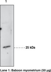
Prostaglandin D synthase (PGDS) catalyzes the isomerization of PGH2 to produce PGD2. PGD2 induces sleep, regulates nociception, inhibits platelet aggregation, and acts as an allergic mediator. Two distinct types of PGDS have been identified, namely the lipocalin type enzyme (β-trace) and the hematopoietic enzyme.1,2,3 Lipocalin type PGDS is localized in the central nervous system and male genital organs of various mammals and the human heart. This enzyme has been identified as β-trace, which is a major protein in human cerebrospinal fluid.2,4 Hematopoietic PGDS is widely distributed in the peripheral tissues and is localized in the antigen-presenting cells, mast cells, and megakaryocytes.1 This enzyme, which requires glutathione for activity, belongs to the sigma-class of glutathione-S-transferases and is approximately 23 kDa in size.5
1
Urade, Y., Watanabe, K., and Hayaishi, O. Prostaglandin D, E, and F synthases. J Lipid Mediat Cell Signal 12 257-273 (1995).
2
Toh, H., Kubodera, H., Nakajima, N., et al. Glutathione-independent prostaglandin D synthase as a lead molecule for designing new functional proteins. Protein Eng 9 1067-1082 (1996).
3
Kanaoka, Y., Ago, H., Inagaki, E., et al. Cloning and crystal structure of hematopoietic prostaglandin D synthase. Cell 90 1085-1095 (1997).
4
Zahn, M., Mäder, A., Schmidt, B., et al. Purification and N-terminal sequence of β-trace, a protein abundant in human cerebrospinal fluid. Neurosci Lett 154 93-95 (1993).
5
Kanaoka, Y., Fujimori, K., Kikuno, R., et al. Structure and chromosomal localization of human and mouse genes for hematopoietic prostaglandin D synthase. Eur J Biochem 267 3315-3322 (2000).
|