| References |
| Stability |
6 months |
| Storage |
4°C |
| Shipping |
Wet ice
in continental US; may vary elsewhere
|
Background Reading
Underhill, D.M., and Ozinsky, A. Phagocytosis of Microbes: Complexity in action. Annu Rev Immunol 20 825-852 (2002).
Beletski, A., Cooper, M., Sriraman, P., et al. High-throughput phagocytosis assay utilizing a pH-sensitive fluorescent dye. Biotechniques 39 894-897 (2005).
| Size |
Global Purchasing |
| 1 ea |
|
Description
Phagocytosis is a very important physiological process which is characterized by the ingestion of foreign particles and killing of microorganisms by phagocytic leukocytes (granulocytes, monocytes, and macrophages). It provides a first line of host defense against potential pathogens. Phagocytosis is used as a model of microbe-innate immune interactions, resulting in increased understanding of the consequences of these interactions.1 Phagocytosis involves a complex series of events including cytoskeletal rearrangement, alterations in membrane trafficking, activation of microbial killing mechanisms, production of pro- and anti-inflammatory cytokines and chemokines, activation of apoptosis, and production of molecules required for efficient antigen presentation to the adaptive immune system. Aberrant phagocytosis has been implicated in several pathological conditions, such as Multiple Sclerosis, Alzheimer’s disease, and atherosclerosis. Deficiencies in phagocytosis cause severe and recurrent bacterial and fungal infections in affected individuals.2
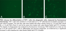
There are a variety of assays available to study the regulation and mechanism of phagocytosis in vitro. Over time, these assays have changed in nature. There were assays employing microscopy to directly visualize engulfed particles, spectrophotometric evaluation of phagocytized paraffin droplets containing dye, or scintillation counting of radiolabeled bacteria. More recently developed assays use fluorescently-opsonized latex beads or fluorescently-labeled bacteria followed by flow cytometric analysis to assess the degree of phagocytosis. The flow cytometric assay has an advantage of rapid analysis of thousands of cells and quantification of internalized particle density for each analyzed cell. Thus, the degree of phagocytosis from cells treated with different compounds can be compared quantifiably. Cayman’s Phagocytosis Assay Kit (IgG FITC) employs latex beads coated with fluorescently-labeled rabbit-IgG as a probe for the identification of factors regulating the phagocytic process in vitro. The engulfed fluorescent-beads can be detected using a microscope capable of measuring excitation and emission at 485 and 535 nm, respectively, or be analyzed by a flow cytometer capable of excitation at 488 nm.
1
Underhill, D.M., and Ozinsky, A. Phagocytosis of Microbes: Complexity in action. Annu Rev Immunol 20 825-852 (2002).
2
Beletski, A., Cooper, M., Sriraman, P., et al. High-throughput phagocytosis assay utilizing a pH-sensitive fluorescent dye. Biotechniques 39 894-897 (2005).
|


